Home
/ Shoulder Joint Anatomy Diagram Easy - 13 Best Shoulder Joint Anatomy Ideas Shoulder Joint Shoulder Joint Anatomy Anatomy _ The human shoulder is the most mobile joint in the body.
Shoulder Joint Anatomy Diagram Easy - 13 Best Shoulder Joint Anatomy Ideas Shoulder Joint Shoulder Joint Anatomy Anatomy _ The human shoulder is the most mobile joint in the body.
Shoulder Joint Anatomy Diagram Easy - 13 Best Shoulder Joint Anatomy Ideas Shoulder Joint Shoulder Joint Anatomy Anatomy _ The human shoulder is the most mobile joint in the body.. Posted on december 13, 2018december 12, 2018. Robin smithuis and henk jan van der woude. Static stabilizing structures include the osseous articular anatomy and joint congruity, the glenoid labrum, the glenohumeral ligaments, joint capsule, and. A patient's guide to shoulder anatomy. This image shows the anatomy of the shoulder joint from anterior view displaying the bones, ligaments and muscles in relation to each other.
Furthermore, glenohumeral joint and its injuries, rotator cuff in conjunction with the acromioclavicular injuries and. A patient's guide to shoulder anatomy. • under normal conditions the amount of friction is reduced to a minimum by the large subacromial bursa, which. Shoulder joint is the most mobile joint of the human body. The shoulder joint is formed where the humerus (upper arm bone) fits into the scapula.
Basic Anatomy Of The Shoulder Acro Physical Therapy Fitness from images.squarespace-cdn.com Human kidney anatomy_easy steps to draw. Static stabilizing structures include the osseous articular anatomy and joint congruity, the glenoid labrum, the glenohumeral ligaments, joint capsule, and. 8 name the arteries and the nerves that supply shoulder joint. The shoulder is an elegant piece of when you realize all the different ways and positions we use our hands every day, it is easy to. Just remember the articulating surfaces. Dislocation of the shoulder is extremely painful and may require surgical repair or even cause permanent damage. The shoulder joint (glenohumeral joint) is a ball and socket joint between the scapula and the humerus. Erythrocyte sedimentation rate (esr) by shabab ali 21392 views.
Chronic or acute wear and tear on the.
The shoulder joint (glenohumeral joint) is a ball and socket joint between the scapula and the humerus. A patient's guide to shoulder anatomy. Chronic or acute wear and tear on the. In common usage, shoulder joint mostly refers to the glenohumeral joint, the major joint of the shoulder but can also include acromioclavicular joint. The human shoulder is the most mobile joint in the body. This incongruent bony anatomy allows for the wide range of movement available at the shoulder joint but is also the reason for the lack of joint stability. Learn vocabulary, terms and more with flashcards, games and other study tools. The shoulder joint by quan fu gan 69549 views. Glenohumeral joint acromioclavicular joint sternoclavicular joint scapulothoracic junction. Shoulder joint of human body anatomy infographic diagram with all parts including bones ligaments muscles bursa cavity capsule cartilage membrane for medical science education and health care. How to draw heart diagram in exams ? The glenohumeral joint (shoulder joint) is a synovial ball and socket articulation anatomy ▶ upper limb ▶ joints ▶ shoulder joint (glenohumeral joint). This diagram here just shows the joint capsule itself.
All about the shoulder muscles. A patient's guide to shoulder anatomy. Robin smithuis and henk jan van der woude. Learn vocabulary, terms and more with flashcards, games and other study tools. Furthermore, glenohumeral joint and its injuries, rotator cuff in conjunction with the acromioclavicular injuries and.
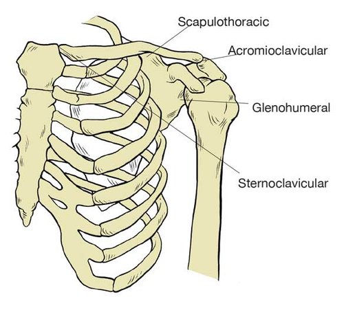
Shoulder Joint Royalty Free Vector Image Vectorstock from cdn1.vectorstock.com Static stabilizing structures include the osseous articular anatomy and joint congruity, the glenoid labrum, the glenohumeral ligaments, joint capsule, and. The shoulder is actually composed of four joints, namely glenohumeral joint, acromioclavicular joint, sternoclavicular joint and scapulothoracic joint. You can see it enclosing the glenohumeral joint and you can see its attachment on the anatomical neck that's the shoulder joint. Dislocation of the shoulder is extremely painful and may require surgical repair or even cause permanent damage. A patient's guide to shoulder anatomy. Shoulder joint of human body anatomy infographic diagram with all parts including bones ligaments muscles bursa cavity capsule cartilage membrane for medical science education and health care. The glenohumeral joint (shoulder joint) is a synovial ball and socket articulation anatomy ▶ upper limb ▶ joints ▶ shoulder joint (glenohumeral joint). Coracoclavicular ligament 3 shoulder joint anatomy.
The glenohumearal joint has a greater range of motion than any other joint in the body.
Human kidney anatomy_easy steps to draw. Diagram of the human shoulder joint, back view. Describe the structure of the shoulder should begin with bone parts that include: Normal anatomy, variants and checklist. Various types of injuries and degenerative conditions can cause the shoulder to become painful. The left shoulder and acromioclavicular joints, and the proper ligaments of the scapula. Three bones come together at the shoulder joint. • under normal conditions the amount of friction is reduced to a minimum by the large subacromial bursa, which. Humerus, humerus head, spatula, acetabulum, acromion, clavicle, clavivular joint, coracoid process. Shoulder joint is the most mobile joint of the human body. 8 name the arteries and the nerves that supply shoulder joint. All about the shoulder muscles. This diagram here just shows the joint capsule itself.
Static stabilizing structures include the osseous articular anatomy and joint congruity, the glenoid labrum, the glenohumeral ligaments, joint capsule, and. Webmd's shoulder anatomy page provides an image of the parts of the shoulder and describes its function, shoulder problems, and more. Glenohumeral joint acromioclavicular joint sternoclavicular joint scapulothoracic junction. Erythrocyte sedimentation rate (esr) by shabab ali 21392 views. Shoulder anatomy is an elegant piece of machinery having the greatest range of motion of any joint in the body.
Anatomy Of The Human Shoulder Joint from www.verywellhealth.com 8 name the arteries and the nerves that supply shoulder joint. In common usage, shoulder joint mostly refers to the glenohumeral joint, the major joint of the shoulder but can also include acromioclavicular joint. Due to the tension by the anterior band of the inferior ghl labral teras will be easier to detect. Three bones come together at the shoulder joint. Shoulder injections injections to the shoulder can be performed either for diagnostic purposes or for aspiration of joint fluid. Just remember the articulating surfaces. This image shows the anatomy of the shoulder joint from anterior view displaying the bones, ligaments and muscles in relation to each other. This diagram here just shows the joint capsule itself.
• during abduction of the shoulder joint, the supraspinatus tendon is exposed to friction against the acromion.
Normal anatomy, variants and checklist. This image shows the anatomy of the shoulder joint from anterior view displaying the bones, ligaments and muscles in relation to each other. Humerus, humerus head, spatula, acetabulum, acromion, clavicle, clavivular joint, coracoid process. This diagram here just shows the joint capsule itself. In common usage, shoulder joint mostly refers to the glenohumeral joint, the major joint of the shoulder but can also include acromioclavicular joint. The shoulder is one of the largest and most complex joints in the body. The shoulder joint is vulnerable to dislocations from sudden jerks of the arm, especially in children before strong muscles have developed. Static stabilizing structures include the osseous articular anatomy and joint congruity, the glenoid labrum, the glenohumeral ligaments, joint capsule, and. This incongruent bony anatomy allows for the wide range of movement available at the shoulder joint but is also the reason for the lack of joint stability. Shoulder joint of human body anatomy infographic diagram with all parts including bones ligaments muscles bursa cavity capsule cartilage membrane for medical science education and health care. The shoulder joint is formed where the humerus (upper arm bone) fits into the scapula. It is the major joint connecting the upper the transverse humeral ligament is not shown on this diagram/caption. All about the shoulder muscles.
Furthermore, glenohumeral joint and its injuries, rotator cuff in conjunction with the acromioclavicular injuries and shoulder anatomy diagram. In common usage, shoulder joint mostly refers to the glenohumeral joint, the major joint of the shoulder but can also include acromioclavicular joint.

/shoulder_pain_medreview-01-5c3b9f8546e0fb0001bdeaaa-d0a4923b7a3d441fb12d992c454a8ca7.png)